

2.5 细胞内源致病因子的检测
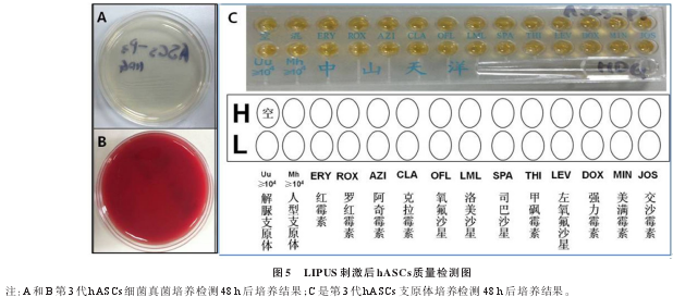
刺激后的第 3代 hASCs 细胞进行实时定量PCR检测人源特定病毒包括HBV、HCV、CMV进行DNA表达量,检测结果均为阴性(<5.00E+02 copy)。
2.6 内毒素检测结果
刺激后的第 3 代 hASCs细胞通过“鲎试剂灵敏度复核试验”法检测细胞培养上清中内毒素含量,结果均为阴性,λ=0.25 EU/mL,浓度值均<2.83 EU/mL.
2.7 革兰氏染色结果
刺激后的第 3 代 hASCs细胞染色,油镜下观察,结果均无革兰氏细菌生长。
3 讨 论
干细胞应用于临床治疗和再生医学的潜能已被干细胞研究者们广泛关注。而一个治疗的疗程至少需要1×10⁷个细胞,但从患者、捐赠者身上无法获得足够的hASCs,限制了其应用[15].有学者探索通过改变各种培养条件,如不同的细胞因子和生长因子,促进体内来源的ASCs体外扩增,目前尚未取得临床相关结论。Jie Chen团队研究发现LIPUS不仅促进人类脐带和骨髓来源的间充质干细胞(MSC)的增长和集落形成,而且促进造血祖干细胞(HSPC)增殖和分化[12],表明可利用LIPUS来获得更多MSC以用于临床[12,16].
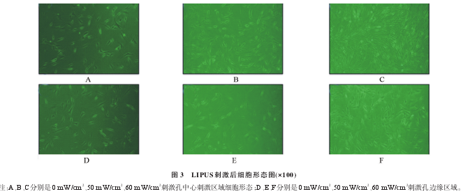
通过研究,笔者发现强度为60 mW/cm2的LIPUS有效促进hASCs的扩增,与对照组比较细胞数量增加约17%,细胞倍增时间缩短为对照组的0.875倍。需要的细胞数量少,且刺激时间短,是有效扩增hASCs的方法之一。本实验存在的不足是未进行更低频率刺激和延长刺激时间研究。
研究显示,LIPUS 不仅能促进 HSPC 亦能促进hASCs 有效增殖。LIPUS 刺激不改变干细胞培养条件,只改变细胞微环境或能量转换使干细胞增殖,避免外源细胞因子和生长因子等影响。因此,我们的研究结果将可以为干细胞体外快速增殖和产业化提供新途径。下一步我们将验证其增殖的安全性。
参 考 文 献
[1] Qin L, Fok P, Lu H, et al. Low intensity pulsed ultrasound increasesthe matrix hardness of the healing tissues at bone-tendon insertion-apartial patellectomy model in rabbits [J]. Clin Biomech (Bristol,Avon), 2006, 21(4): 387-394.
[2] He R, Zhou W, Zhang Y, et al. Combination of low-intensity pulsedultrasound and C3H10T1/2 cells promotes bone- defect healing [J].Int Orthop, 2015, 39(11): 2181-2189.
[3] Leung KS, Cheung WH, Zhang C, et al. Low intensity pulsed ultra-sound stimulates osteogenic activity of human periosteal cells [J].Clin Orthop Relat Res, 2004, (418): 253-259.
[4] Hanawa K, Ito K, Aizawa K, et al. Low-intensity pulsed ultrasoundinduces angiogenesis and ameliorates left ventricular dysfunction ina porcine model of chronic myocardial ischemia [J]. PLoS One,2014, 9(8): e104863.
[5] Bernal A, Pérez LM, De Lucas B, et al. Low-Intensity pulsed ultra-sound improves the functional properties of cardiac mesoangioblasts[J]. Stem Cell Rev, 2015, 11(6): 852-865.
[6] Padilla F, Puts R, Vico L, et al. Stimulation of bone repair with ultra-sound: a review of the possible mechanic effects [J]. Ultrasonics,2014, 54(5): 1125-1145.
[7] dos Santos L, Santos AA, Gon?alves GA, et al. Bone marrow cell ther-apy prevents infarct expansion and improves border zone remodelingafter coronary occlusion in rats [J]. Int J Cardiol, 2010, 145(1): 34-39.
[8] Mummery CL, Davis RP, Krieger JE. Challenges in using stem cellsfor cardiac repair [J]. Sci Transl Med, 2010, 2(27): 27ps17.
[9] Zuk PA, Zhu M, Ashjian P, et al. Human adipose tissue is a source ofmultipotent stem cells [J]. Mol Biol Cell, 2002, 13(12): 4279-4295.
[10] Lee RH, Kim B, Choi I, et al. Characterization and expression analy-sis of mesenchymal stem cells from human bone marrow and adiposetissue [J]. Cell Physiol Biochem, 2004, 14(4-6): 311-24.
[11] Kern S, Eichler H, Stoeve J, et al. Comparative analysis of mesenchy-mal stem cells from bone marrow, umbilical cord blood, or adiposetissue [J]. Stem Cells, 2006, 24(5): 1294-1301.
[12] Xu P, Gul- Uludag H, Ang WT, et al. Low- intensity pulsed ultra-sound-mediated stimulation of hematopoietic stem/progenitor cell vi-ability, proliferation and differentiation in vitro [J]. Biotechnol Lett,2012, 34(10): 1965-1973.
[13] 国家卫生计生委办公厅, 食品药品监管总局办公厅。 卫计委印发干细胞制剂质量控制及临床前研究指导原则(试行)通知[S]. 2015.http://www.nhfpc.gov.cn/qjjys/s3581/201508/15d0dcf66b734f338c31f67477136cef.shtml.
[14] 国家药典委员会。 中华人民共和国药典[M]. 3 版。 北京: 中国医药科技出版社, 2010, 附录: IX B-XIII D.
[15] Bernal A, Pérez LM, De Lucas B, et al. Low- Intensity rulsed ultra-sound improves the functional properties of cardiac mesoangioblasts[J]. Stem Cell Rev, 2015, 11(6): 852-865.
[16] Xing JZ, Yang X, Xu P, et al. Ultrasound -enhanced monoclonal anti-body production [J]. Ultrasound Med Biol, 2012, 38(11): 1949-1957.


